Isoelectric focusing (IEF) is mainly used for the determination of pI values. Due to its specific selectivity IEF can also be applied for the challenging separations like isoform antibodies.
In comparison to the other separation techniques IEF offers more points for optimization like the nature and concentration of the ampholyte, concentration of catholyte and anolyte, respectively as well as the dimension of the capillary, its coating and the kind of mobilization.
cIEF of a peptide mixture, without IEF filter
- Mode: cIEF
- Ampholyte: 3-10
- Capillary: µSil, 50 µm ID, 32,5 in total
- Separation: 30 kV
- Mobilization: chemically after 5 min with 350 mM acetic acid
- Detection: direkt UV, 275 nm, without filter
- Catholyte: NaOH
- Anolyte: Phosphoric acid
- Description: The standard solution contains peptide markers as well as the ampholyte with gel and urea. Calibration is possible due to the standard markers.
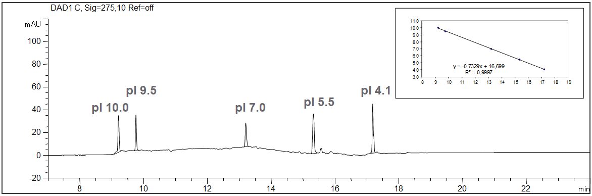
IEF peptide standard with Calibration
Download: IEF-Peptidmarker
cIEF of a monoclonal antibody mAK, influence of the ampholyte
- Mode: cIEF
- Ampholyte: 3-10
- Capillary: µSil, 50 µm ID, 32,5 in total
- Separation: 30 kV
- Mobilization: chemically after 5 min with 350 mM acetic acid
- Detection: direkt UV, 275 nm, without filter
- Catholyte: NaOH
- Anolyte: Phosphoric acid
- Description: The PDF shows the influence of the ampholyte on the separation. Good separation of the isoforms is already achieved with the broadband ampholyte 3-10. Due to the limitation of the pI range to 5-8 resolution can be increased drastically. The sample solution was prepared including the peptide markers and the particular ampholyte with gel and urea. The exact determination of the pI of the separated isoforms of mAK is possible
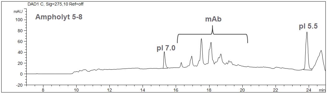
IEF of a monoclonal antibody – Comparison with different ampholytes
Download: IEF-Ampholyte
cIEF of a peptide mixture with IEF filter
- Mode: cIEF
- Ampholyte: 3-10
- Capillary: µSil, 50 µm ID, 32,5 in total
- Separation: 30 kV
- Mobilization: chemically after 6 min with 350 mM acetic acid
- Detection: direkt UV, 280 nm, with filter (Agilent G7100-68750)
- Catholyte: NaOH
- Anolyte: Phosphoric acid
- Description: Whereas the pictures given above showed separation made without filter the example here shows a significant improvement of the noise as well as the sharpness of the peaks due to the installation of the filter. The filter can easily and without the use of tools be inserted in front of the DAD detector. The PDF also show the typical shape of the current during the cIEF separation and a calibration.
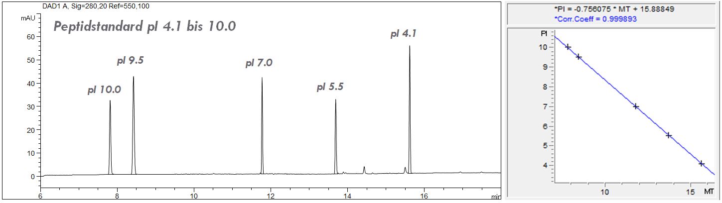
cIEF of a peptide mixture with filter
Download: IEF-Peptidmarker-mit-filter
cIEF of a monoclonal antibody IgG1 with IEF filter
- Mode: cIEF
- Ampholyte: 3-10
- Capillary: µSil, 50 µm ID, 32,5 in total
- Separation: 30 kV
- Mobilization: chemically after 6 min with 350 mM acetic acid
- Detection: direkt UV, 280 nm, with filter (Agilent G7100-68750)
- Catholyte: NaOH
- Anolyte: Phosphoric acid
- Description: Under the given conditions the isoforms of the monoclonal antibody typ IgG1 are clearly separated and the determination of their pIs was possible. The PDF shows the separation as well as the pI values and the calibration curve.
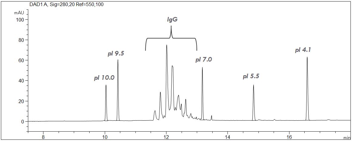
cIEF of a monoclonal antibody IgG1 with filter
Download: IEF-mAb-mit-Filter